'Use of CT imaging to identify osteopenia and fractures
in a 2 year old Miniature Schnauzer'
Patient:
Miniature Schnauzer, Female, 2 years old
Clinical Background:
• Intermittent history of abnormal lameness lasting 6-8 weeks
• Waxing and waning lameness affecting right and left forelimb
• Concern about radio ulnar ischaemic necrosis on radiography
• Previously referred to boarded neurologist who did not note any abnormality on exam
Clinical Questions:
• Have any abnormalities been seen in the patients forelimbs to explain the history of lameness?
• Have any abnormalities been noted to be affecting the chest?
Clinical Plan:
• CT Chest Pre/Post Contrast
• CT Forelimbs Pre/Post Contrast
CT Scans Performed:
• Patient was positioned prone with forelimbs out stretched in front
• AP and lateral survey done from tip of paws down to bottom of chest
• Pre contrast scans performed of chest and forelimbs
• Post contrast scans performed of chest (arterial) and forelimbs
Clinical Diagnosis:
The CT Scan report was as follows: (see figure 1 below)
-
Generalised decreased bone attenuation of the veterbral bodies is evident, accompanied by cortical thinning
-
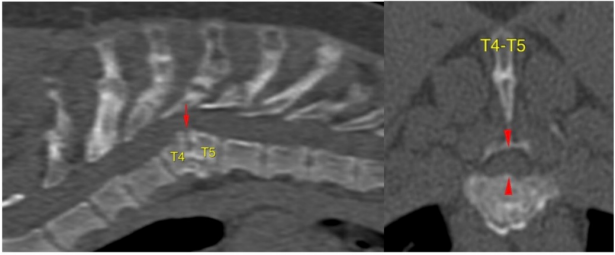
The thoracic vertebral bodies T4 and T5 are shortened and malformed, less pronounced also T2.
-
The intervertebral disc space is narrowed between T4-T5
-
Partial bridging ventrolateral spondylosis is present at T4-T5
-
Malalignment/dorsal kinking can be demonstrated between T4-T5, resulting in mild dorsoventral flattening/stenosis of the vertebral canal and mild spinal cord compression respectively, slightly more pronounced to the right side
Figure 1
-
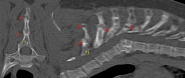
Fractures of the spinous process of the cranial thoracic spine (T1-T5) can be demonstrated, which are partially remodelled/chronic or hypertrophic and non-union healing (see figure 2 below)
Figure 2
-
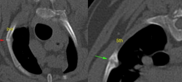
Furthermore fractures of multiple right and left ribs can be identified (2-5th and few caudal right ribs 3-6th) left ribs), partially with kinked malalignment or hypertrophic, non union healing (see figure 3 below)
-
The hypertrophic bone of several rib fractures protrudes mildly into the thoracic cavity
Figure 3
-
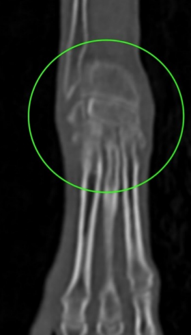
Similar to the cervicothoracic spine, generalised decreased bone attenuation of the appendicular skeleton of both forelimbs cannot be demonstrated, accompanied by cortical thinning and poor or even absent mineralisation of the cortices of the epiphyses and the subchondral bone, most pronounced at the level of carpus, elbows and to a lesser extent of shoulders (see figures 4 & 5 below)
-
No cortical bone can be seen along the radial incisura or around the medial coronoid process of both elbows
-
The trabecular bone is generally poorly mineralised
-
The joint space appears in many areas collapsed
-
The long bone cortices are relatively dense
-
There are mild degenerative changes, mainly in both carpi
-
The scapula appears relative thin and mildly irregular bilaterally
Figure 4
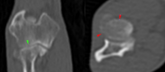
Figure 5
Clinical Results:
-
Osteopenia with partially severe cortical thinning at the level of the epiphysis and subchondral bone can be identified in both forelimbs, less pronounced cervicothoracic spine
-
A metabolic bone disorder is the most likely cause for the bilateral symmetrical changes in both forelimbs and bilateral forelimb lameness
Burgess Diagnostics Senior Radiographer: Sam Kolodziejczak
